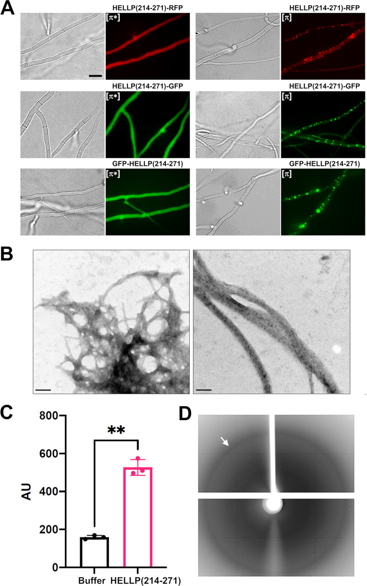
FIG 2.
Characterization of the PP motif of P. anserina. (A) PP motif region of HELLP behaves as a prion-forming domain in vivo, as shown on micrographs of P. anserina strains expressing different molecular fusions [HELLP(214-271-RFP)/GFP and GFP-HELLP(214-271)], as indicated above each micrograph. Bar, 5 μm. Transformants initially present a diffuse fluorescence in the nonprion state designated [π*] (left) and systematically acquire dot-like fluorescent aggregates after contact with a strain already expressing the infectious prion state designated [π] (right). (B) The PP motif forms fibrils in vitro, as shown on an electron micrograph of negatively stained HELLP(214-271) fibrils (bar, 100 nm). (C) ThT-fluorescence signal of HELLP(214-271) fibrils. HELLP(214-271) fibrils and a buffer control were incubated with ThT, and fluorescence was measured (excitation wavelength, 440 nm; emission wavelength, 480 nm). **, P = 0.0028 (Welch’s test). (D) The X-ray diffraction pattern of unoriented HELLP(214-271) fibrils is given, and the reflection at 4.7 Å is marked by an arrow.

