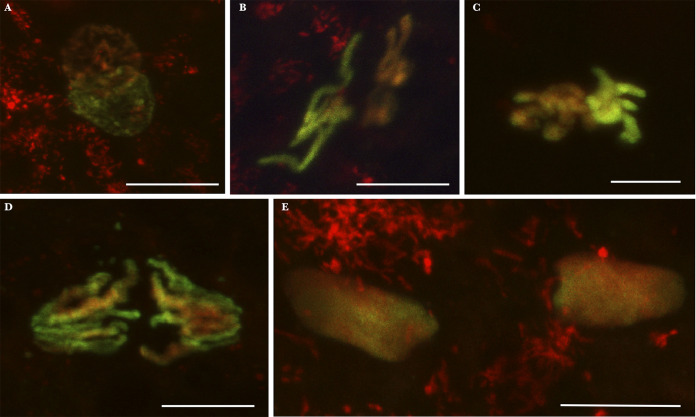
FIG 2.
Culex pipiens embryos from INTER-INTER crosses exhibiting normal first division. Paternal chromatin appears in green/yellow (acetylated histone H4 labeling is dominant), and maternal chromatin appears in red (propidium iodide labeling is dominant). These embryos have been collected and fixed 30 min to 1 h postoviposition. (A) Apposition of maternal and paternal pronuclei; (B) chromatin under condensation; (C) condensed chromatin; (D) first mitotic division anaphase; (E) two nuclei following the first division. Scale bar represents 10 μm.

