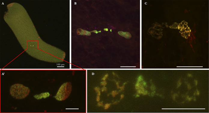
FIG 3.
Culex pipiens embryos from INTER-INTER crosses exhibiting CI in first division. Paternal chromatin appears in green/yellow (acetylated histone H4 labeling is dominant), and maternal chromatin appears in red (propidium iodide labeling is dominant). These embryos have been collected and fixed in the first hour postoviposition. (A) Global view of a C. pipiens embryo undergoing a first mitotic division. (A′) Magnification of panel A showing paternal chromatin failed to segregate properly and form a chromatin spot between segregated nuclei. (B, C, and D) Other kinds of failed first divisions observed. Confocal stacks were obtained on embryos from several INTER-INTER crosses. Scale bar represents 10 μm.

