
FIGURE 1.
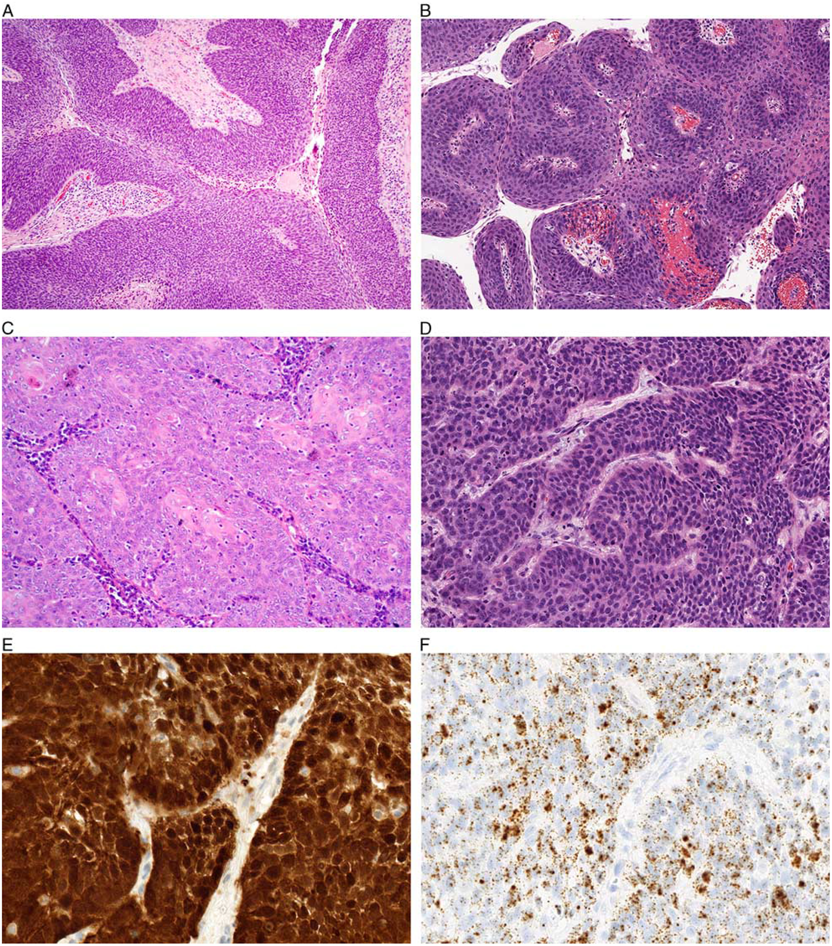
The majority of nonoropharyngeal HPV-positive SCCs demonstrated a nonkeratinizing morphology with nests and lobules of somewhat syncytial basaloid cells with minimal desmoplastic stroma (A, oral cavity). Although no tumors were entirely exophytic, a subset demonstrated papillary architecture (B, larynx). The tumors showed minimal keratinization in a central or peripheral distribution (C, larynx). While cells had a high nuclear-cytoplasmic ratio, nuclei were oval and relatively uniform with only focal pleomorphism (D, larynx). These nonkeratinizing tumors were diffusely positive for p16 (E, larynx) and HPV RNA ISH (F, larynx).