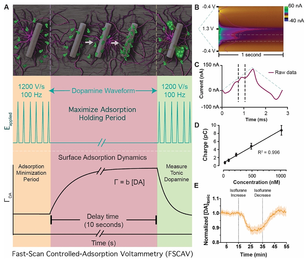
Fig. 2.

Fast-scan controlled-adsorption voltammetry. (A, Left) Adsorption of dopamine at the electrode surface is minimized during the application of the 100 Hz triangle waveform. (A, middle) The electrode is held at a negative potential for 10 s, during which time dopamine maximally adsorbs to the electrode surface and an adsorption equilibrium is reached. (A, right) The 100 Hz triangle waveform is re-applied, and information rich measurement is made that represents the equilibrium dopamine concentration (i.e., the tonic level). (B) Pseudo-color representation of recorded current and superimposed integration limits (horizontal dashed lines) typically used for dopamine quantification. (C) Current taken from the 12th scan after the holding period is used for tonic dopamine quantification. Superimposed integration limits and applied waveform are shown in dashed lines. (D) Calibration of electrodes in dopamine solutions demonstrates a linear dynamic range over several orders of magnitude. Adapted from Ref. [38] with permission from the Royal Society of Chemistry. (E) Example of tonic dopamine detection data in response to changing levels of the anesthetic isoflurane.
