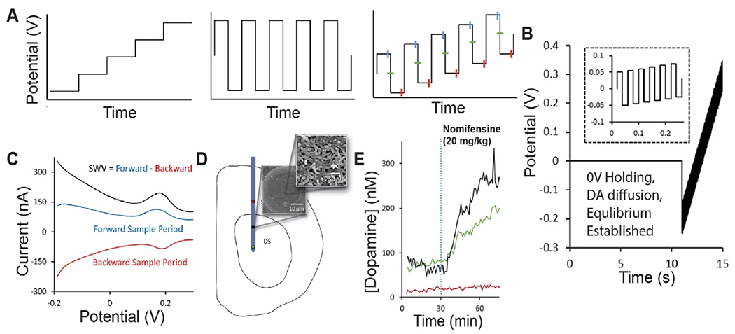
Fig. 4.

PEDOT/CNT-functionalized carbon-fiber microelectrode measurements of tonic dopamine via SWV. (A) The waveform consists of the superposition of a staircase wave and a square wave. Forward current is measured at the end of each anodic holding period, and backward current is measured at the end of each cathodic holding period. (B) An initial 0 V hold followed by a series of anodic and cathodic step and holds (zoom shown in the inset) that transverse the defined potential window is applied. (C) Representative SWV measurement of a 1 μM standard solution of dopamine reveals the SWV current (black) response to be the difference of the forward (blue) and reverse (red) current responses. (D) Schematic of microelectrode array placement in the dorsal striatum (DS) of the rat. Multiple recording sites indicated by red, black, and green circles. SEM image of CNT functionalization on electrode surface. (E) Electrodes located within the DS (green and black) showed clear, nomifensine-dependent DA detection. Adapted with permission from Ref. [43]. Copyright 2019 American Chemical Society.
