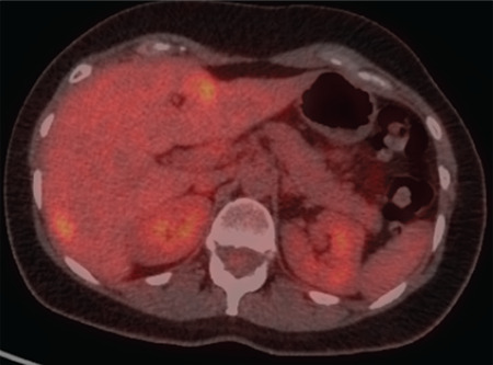
Figure 5.

A baseline PET/CT of a patient with 2 liver metastases from breast cancer, who was treated with RFA. Pre-treatment axial fused slice on the level of liver segments 2 and 7, showing metabolically active liver lesions respectively with SUVmax 5.2 and SUVmax 4.3
PET/CT: Positron emission tomography/computed tomography, RFA: Radiofrequency ablation, SUVmax: Maximum standard uptake value
