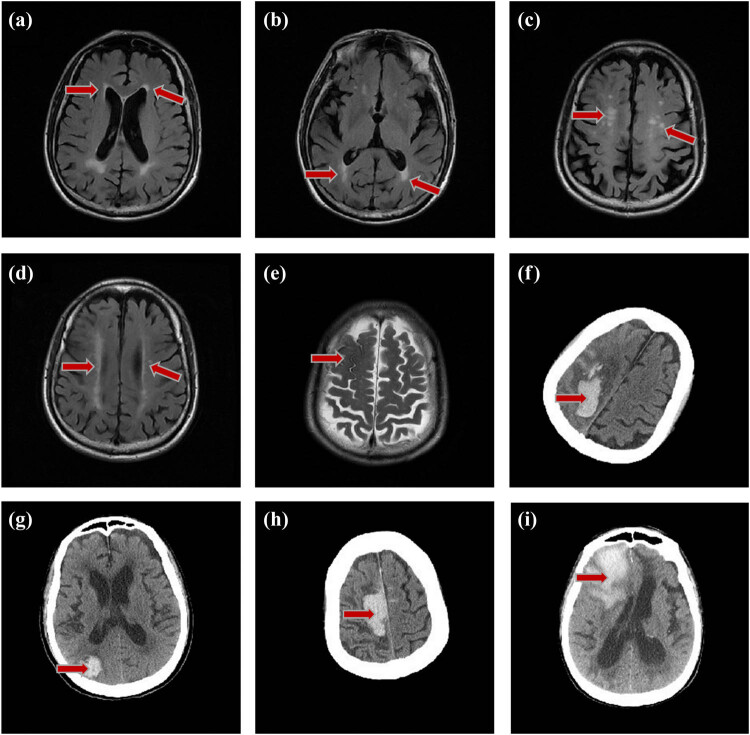
Figure 2.
Distribution of early LA and hemorrhages in case 12. An 86-year-old male patient, occipital prominent LA and subcortical patchy distribution of white matter appeared 3 years before the ICH. (a) Score 1 in the frontal lobe white matter (periventricular 1, deep 0, juxtacortical 0). (b) Score 4 in the occipital lobe white matter (periventricular 3, deep 1, juxtacortical 0). (a and b) FO value = −3 occipital prominent LA. (c) Subcortical multiple punctate LA, without clear periventricular white matter damage. (d) The bilateral white matter lesions were asymmetric, with the right white matter lesions severer. (e) T2 shows old convex subarachnoid hemorrhage in August 2009. (f) Shows that ICH occurred in right parietal-occipital lobe in 2012. (g–i) CT shows recurrent ICH occurred in right occipital lobe, parietal lobe, and frontal lobe in 2014, 2016, and 2018, all on the same side of convex subarachnoid hemorrhage.

