Fig. 2.
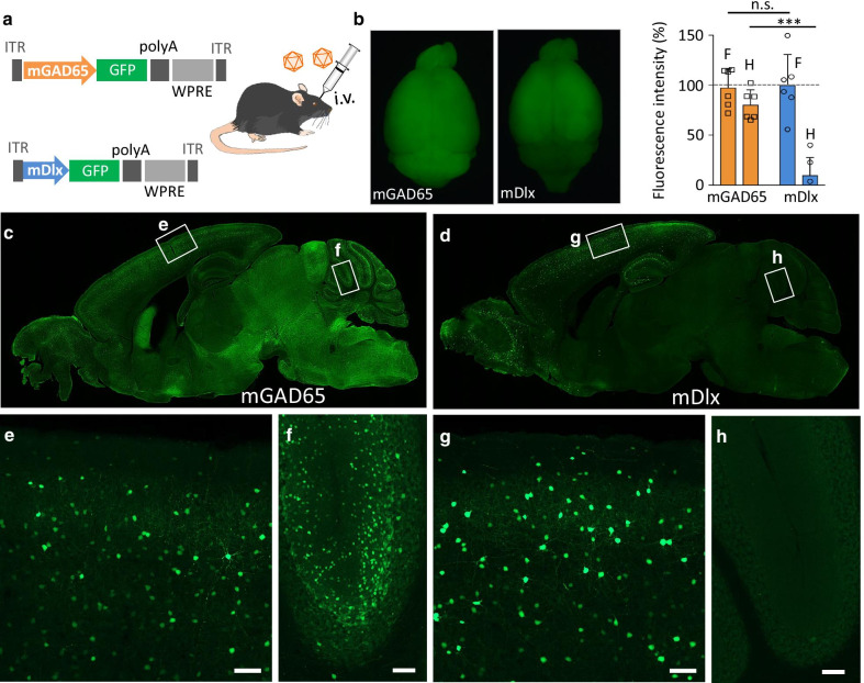
Native GFP of sagittal sections from mice 3 weeks after intravenous injection of AAV vectors. a Schematic showing the expression cassettes of AAV vectors, which express GFP under the control of the mGAD65 promoter or mDlx enhancer. The mice received intravenous injections (i.v.) of the blood–brain barrier (BBB)-penetrating AAV-PHP.B. b Fluorescent stereoscopy of whole brains expressing GFP by the mGAD65 (left) or mDlx enhancer (right). Graph next to the brains shows the relative GFP fluorescent intensities (average ± SEM) in the forebrain (F) and hindbrain (H) transduced by the mGAD65 promoter or mDlx enhancer. Values from respective mice (n = 6 mice in each group) were plotted on each bar graph. ****p < 0.0001 by 2-way ANOVA for repeated measures with Bonferroni’s posthoc test. c, d Sagittal brain sections from mice expressing GFP under the control of the mGAD65 promoter (c) or mDlx enhancer (d). e–h Enlarged GFP fluorescent images of square regions in panels (c and d). Scale bars; 100 μm

