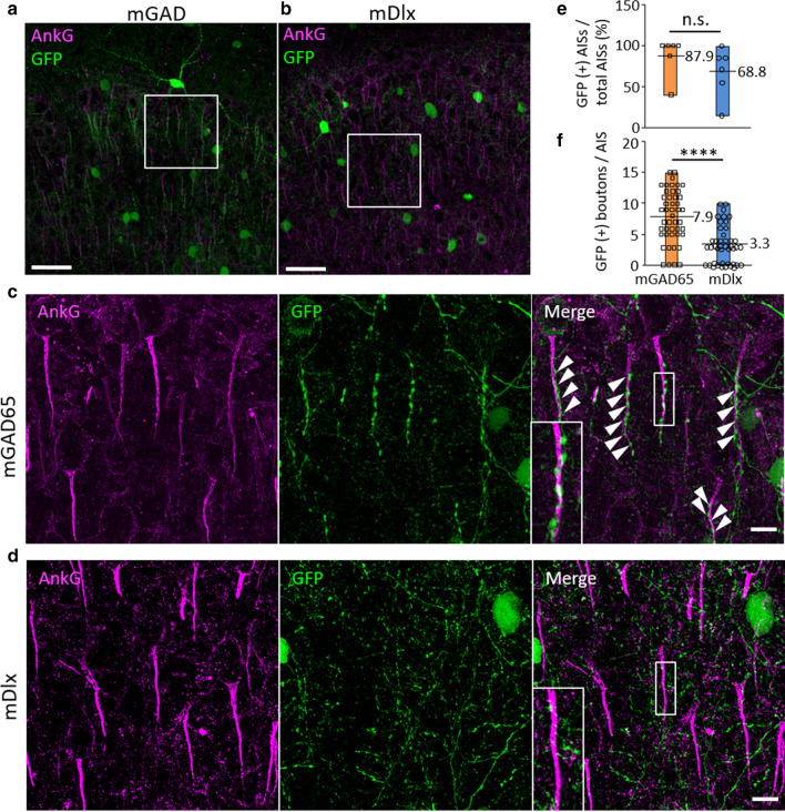
Fig. 5.
GFP labeling of the chandelier cell cartridges by the mGAD65 promoter. Immunohistochemistry of the prefrontal cortex. Sagittal sections from wild-type mice treated with AAV-PHP.B expressing GFP under the control of the mGAD65 promoter a or mDlx enhancer (b) were immunostained for ankyrin-G (magenta). c, d Enlarged images of the square regions in A, B. Note bead-like GFP labeling of chandelier cell synaptic boutons on the axon initial segments (AIS) of excitatory pyramidal neurons (arrowheads). Insets in the upper and lower right panels show the magnification of the boxes. Scale bars; 50 μm (a, b) and 20 μm (c, d). e The ratio of AISs with GFP ( +) boutons to total AISs in the prefrontal cortex. (F) The number of GFP ( +) boutons per AIS in the prefrontal cortex. Values adjacent to the floating boxes in (e, f) are the averages. ****p < 0.0001 on unpaired t-test

