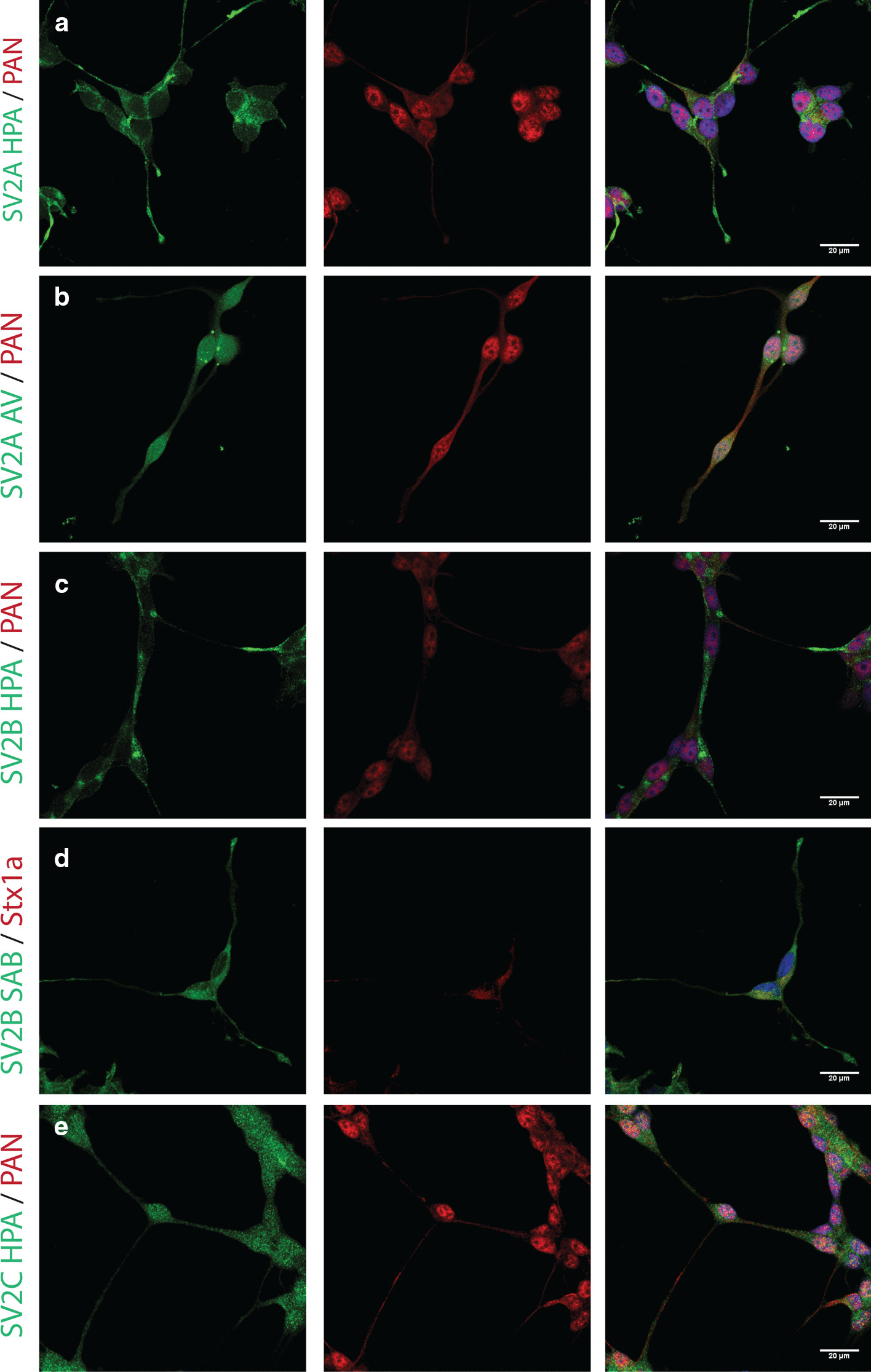
Fig. 1.

Immunocytochemistry of SiMa SV2 proteins. SV2 isoforms were stained in green, Pan neuronal cocktail in red, nuclear staining using DAPI depicted in blue. Images acquired using confocal microscopy, merged images of three stacks. Scale bar 20 µm. Several antibodies were used for the SiMA cell lines. a, b SiMa cells stained for SV2A (green) using two different antibodies and neuronal marked (red) reveal punctuate SV2A staining in processes and around soma. c, d SiMa cells stained for SV2B (green) using two different antibodies and neuronal marker (red) or Stx1a (red) reveal moderate staining in projections. e SiMa cells stained for SV2C (green) and neuronal marked (red) reveal staining in the cell soma
