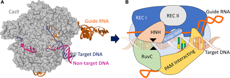
FIGURE 1.
CRISPR/Cas9 structure. (A) X-ray structure of the Streptococcus pyogenes (Sp) CRISPR/Cas9 system (5F9R.pdb) in the pre-activated state (Jiang et al., 2016), created using Mol* (Sehnal et al., 2018). Cas9 (gray) is shown in molecular surface. The guide RNA (orange), the target DNA (dark blue), and non-target DNA (pink) strands are shown as cartoons. (B) A schematic CRISPR/Cas9 ribonucleoprotein structure formed by six domains: Rec I, Rec II, RuvC, HNH, Bridge Helix, and PAM Interacting domain, and guide RNA targeting DNA. The black arrow heads indicate the cut sites from each RuvC and HNH domains. The yellow/green nucleotides represent the PAM sequence.

