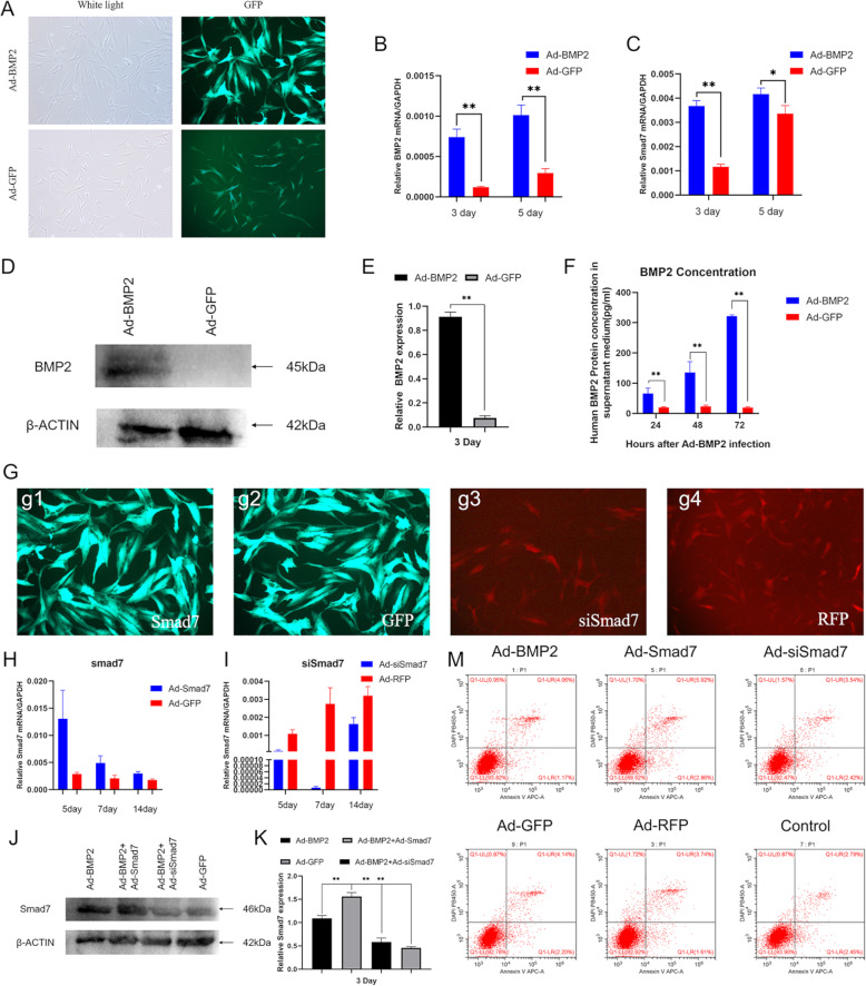
Fig. 2.
Adenovirus-mediated effective and safe transduction of BMP2, Smad7, siSmad7, RFP, and GFP into hSMSCs. a Representative bright field and GFP fluorescence fields were recorded at 24 h after infection (scale bar = 100 μm). b RT-PCR analysis of adenovirus-mediated transgene expression. Total RNA was isolated at 3 and 5 days after infection and subjected to RT-PCR using BMP2 primers. c RT-PCR analysis of Smad7 expression at mRNA level. BMP2 upregulated Smad7 mRNA levels at days 3 and 5 compared to the GFP group. d Western blot analysis of adenovirus-mediated transgene expression. Total cell lysate was collected from the hSMSCs at 72 h after infection and subjected to SDS-PAGE. The transgene expression of BMP2 was probed. Ad-GFP infected cells were used as negative controls. b-actin expression was used as loading controls. e The quantification results of the western blotting assay showed the increased expression level of BMP2. f Quantification of BMP2 protein levels on the medium supernatant by ELISA. Data were collected on 24 h, 48 h, and 72 h after adenovirus transduction. g The recombinant adenovirus Ad-Smad7 (green,g1), Ad-GFP (green,g2), Ad-siSmad7 (red,g3), and Ad-RFP (red,g4) were shown to effectively transfect hSMSCs for 48 h (scale bar = 100 μm). h Ad-Smad7 upregulated the expression of Smad7 at 5, 7, and 14 days. i Ad-siSmad7 silences the expression of Smad7 from 5 to 14 days. All samples were normalized with the house-keeping gene GAPDH. j Western blot analysis for the expression of Smad7 was conducted at day 3 after transduction of indicated recombinant adenoviruses (k), and quantitatively, relative Smad7 expression was analyzed and using b-actin as controls. m hSMSCs were infected with the indicated adenoviral vectors for 3 days. The infected cells were collected and subjected to apoptosis assay. Each experiment was done in triplicate. The data are shown as mean ± SD for triplicate. *P < 0.05, **P < 0.01

