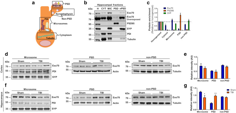
Fig. 4.
Exo70 is redistributed into PSD in the hippocampus of mTBI mice. a Schematic representation of subcellular fractionation proteins. b Example of subcellular fractionation. Hippocampus from two-month-old male mice was fractionated and microsome, PSD, and nonPSD fractions were obtained. 20 µg of protein samples were resolved in a 10% SDS-PAGE and transferred to PVDF membranes. Membranes were incubated with the respective antibodies shown in the figure. Membranes were stripped and tested again with the indicated antibodies. c Proteins distribution were analyzed with densitometric analysis by comparing signal intensity from each fraction with homogenized. Mean values ± SEM are shown. d Cortex and Hippocampus (f) from Sham and mTBI mice were fractionated and analyzed by western blot using Exo70, PDI, Actin, and Tubulin antibodies. PDI/Actin/Tubulin was used as loading controls. 30 µg of protein samples were used. e, g The graph shows the Exo70 densitometric analysis normalized with loading controls. Values represent means ± SEM, n = 3 mice per experimental group. Statistical differences were determined by an unpaired t-test comparing Sham and mTBI. *p < 0.05, **p < 0.01

