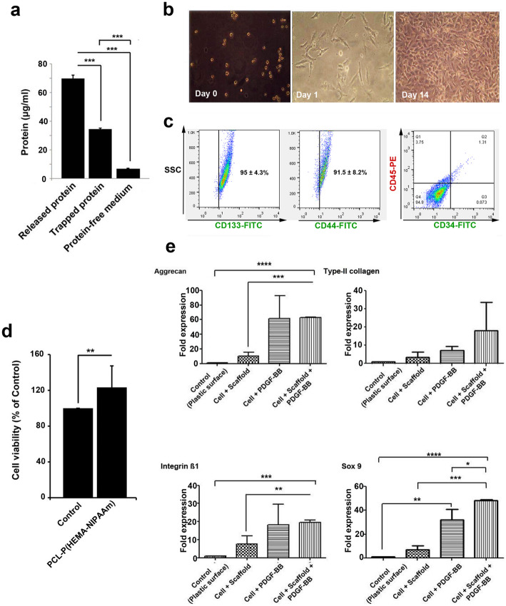
Fig. 4.
Measurements of the total protein contents in the PCL-P(HEMA-NIPAAm) S-2 hydrogel after 2 weeks of incubation with serum-containing medium using a BCA assay as described in the Materials and Methods. ***p < 0.001 (a). Morphological analysis by inverted phase-contrast microscopy (magnification × 100) at a different stages of culture and proliferation (days 0, 1, and 14) (b). Characterization of hASCs (c). Detection of hASC surface marker expression (CD133, CD44, CD34, and CD45) by flow cytometry as described in the Materials and Methods. Viability of hASCs in the PCL-P(HEMA-NIPAAm) S-2 hydrogel. Samples were processed using the cytotoxicity assay after 14 days. **p < 0.01 (d). Real-time RT-PCR analysis of the gene expression profiles in hASCs seeded on the PCL-P(HEMA-NIPAAm) S-2 hydrogel (e). Aggrecan, type-II collagen, SOX9, and integrin β1 expression was analyzed using the 2-ΔΔCT method. GAPDH was used as a housekeeping gene and internal control. *p < 0.050, **p < 0.010, ***p < 0.001, and ****p < 0.0001

