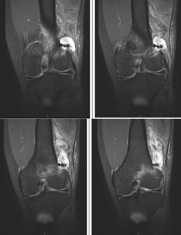
Figure 2.

(a-d) T2-weighted coronal cuts from the MRI of the left knee. The large cystic swelling adjacent to the left lateral distal femur can be seen, as well as the possible communication between the cyst and the femoral tunnel. The Endobutton can be seen, perched on the lateral femoral cortex and possible marrow edema is seen around the femoral tunnel.
