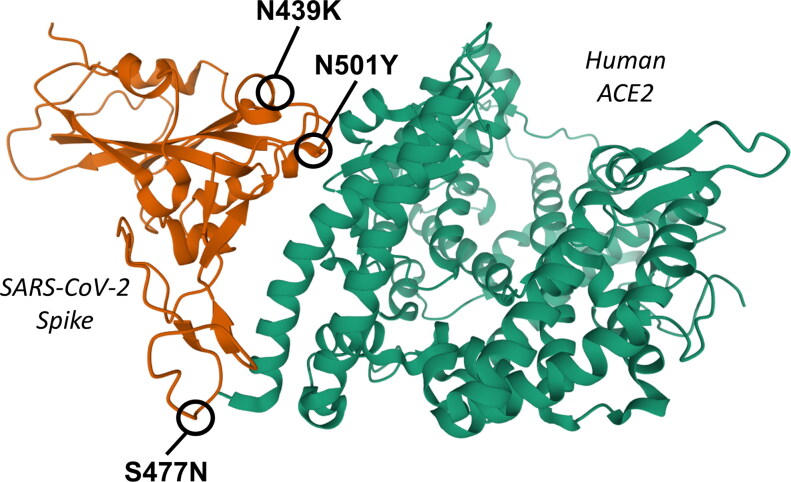
Figure 3.
3 D ribbon representation of the interaction domains of SARS-CoV-2 Spike (left, orange) and human ACE2 (right, green), based on the crystal structure 6LZG deposited on Protein Data Bank and produced by Wang et al. (2020). The positions of the three most frequent Spike mutations in the interacting region (AA 350-550) with a non-zero GBPM score are indicated: N439K, N501Y and S477N.

