
FIGURE 1.
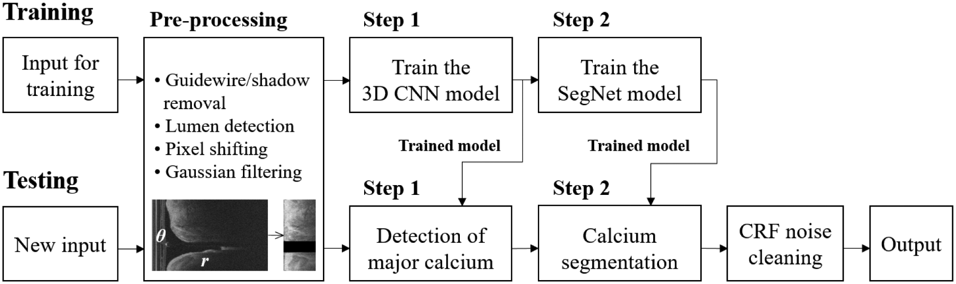
Overall workflow of the proposed two-step deep learning approach for calcium segmentation. Pre-processing is applied to the raw IVOCT image in (r, θ) domain. After pre-processing, the size of the input image is reduced from 968 × 448 to 200 × 448 without any data loss. The trained 3D CNN (step 1) was used to determine the major calcification lesions from the entire pullback. The calcified plaques were segmented using the trained SegNet model (step 2). Classification noises were reduced using a fully connected CRF method. The output label was transformed back to the original size from 200 × 448 to 968 × 448.