
FIGURE 10.
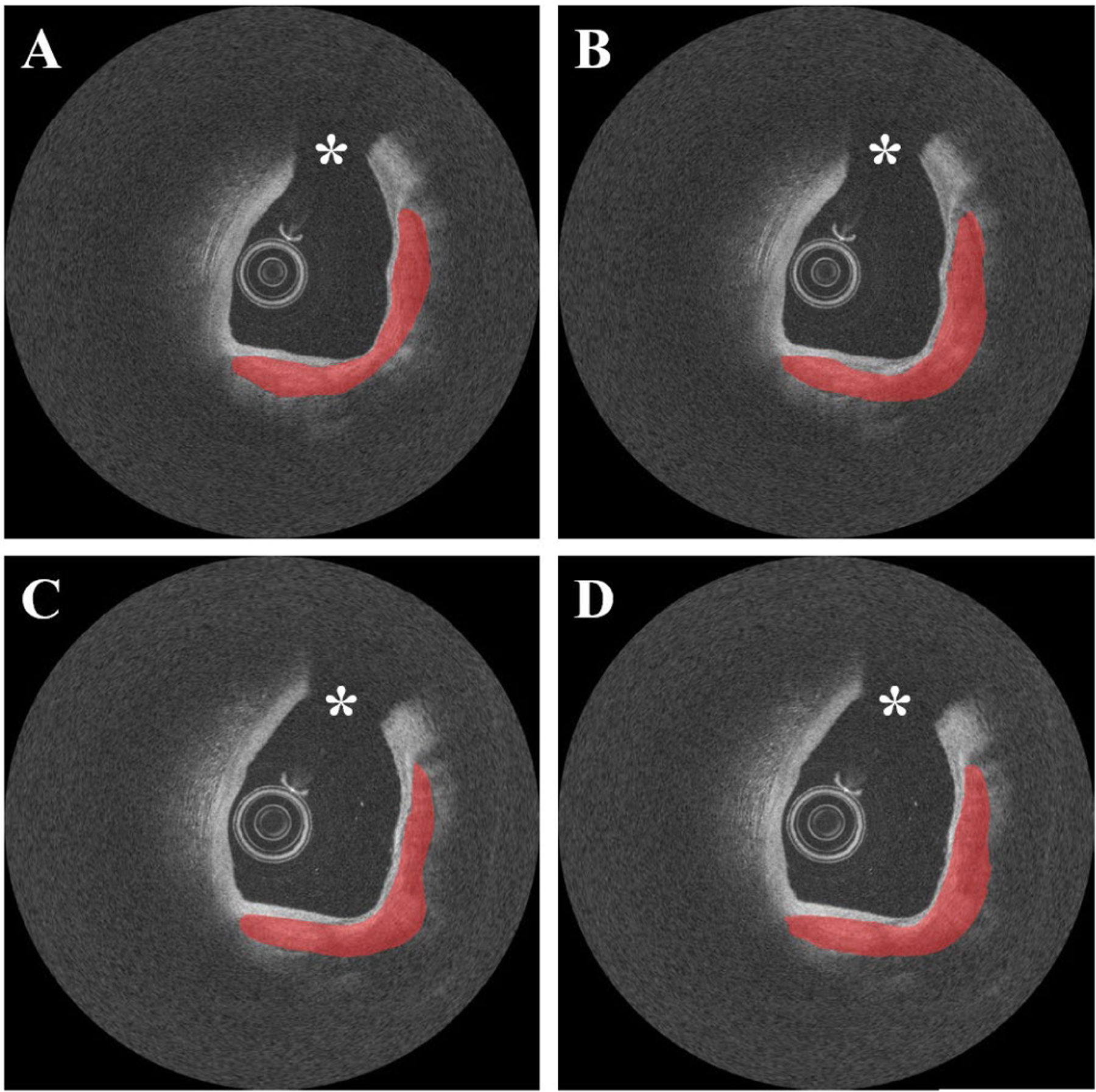
Reproducibility test results on the repeat cadaveric IVOCT pullbacks showing heavily calcified plaques. (A) Ground truth of pullback 1. (B) Prediction of pullback 1. (C) Ground truth of pullback 2. (D) Prediction of pullback 2. Although the cadaveric images have somewhat different intensity profiles, our two-step method produced very similar results on the repeat pullbacks. During the image acquisition, the catheter was placed in the same location. For better visualization, the pullbacks were manually co-registered. Red indicates the calcified plaque, white asterisk (*) indicates the guidewire shadow.