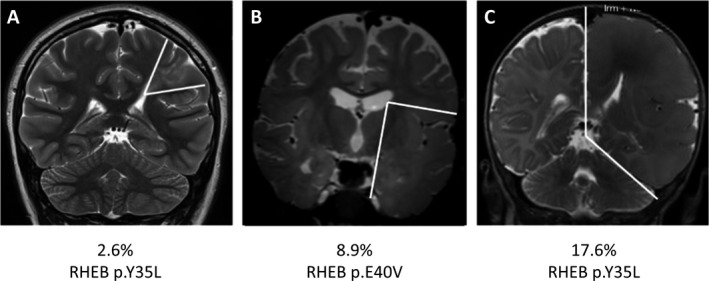
Figure 1.

Correlation between somatic variant load in RHEB and the extent of lesion. Coronal T2 MRI scans showing transmantle FCD in Patient 1 (A) lobar FCD in Patient 2 (B), and HME in Patient 3 (C). The boundaries of the malformations are illustrated by white lines. Somatic variants in RHEB were identified in all three patients, with positive correlation between the variant load and the size of the brain lesion. The corresponding genetic variant and somatic variant load are indicated for each patient.
