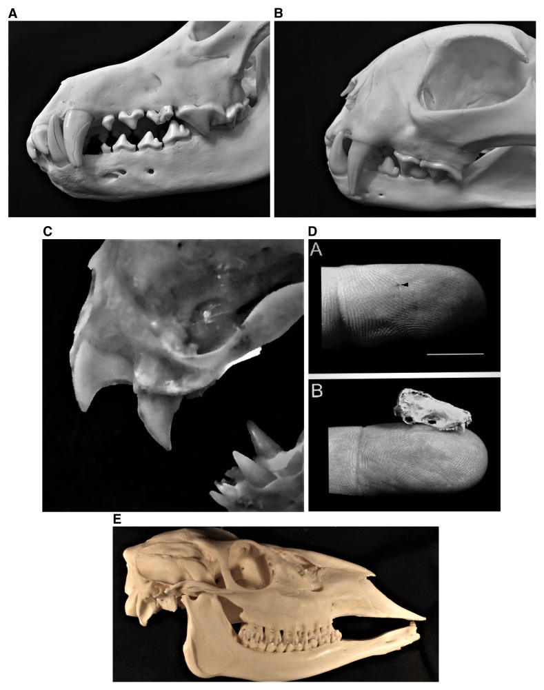
Figure 1. Lyssavirus virions are effectively transmitted transdermally via the saliva into peripheral tissues of a prospective host by the bites of infected mammals, as exemplified by representative mesocarnivores and bats (and in contrast to herbivores).
1A. Close-up of the canines and carnassial teeth of a canid apex predator, the North American gray wolf, Canis lupus. 1B. Close up of the canines and carnassial teeth of the most common rabid wild felid diagnosed within North America, the bobcat, Lynx rufus. 1C. Close up of the specialized incisors and canines of the common vampire bat, Desmodus rotundus. 1D. Example of an insectivorous bat bite. A. Demonstration of a typical small lesion to a finger from an insectivorous bat (bar inset approximately 1 cm). B. Comparison of the skull of an insectivorous bat to a human digit. This figure was reprinted with permission from Elsevier (Jackson AC, Fenton MB. Human rabies and bat bites. The Lancet. 2001. 357:1714)201. 1E. Lateral view of the skull of a representative mammalian herbivore, demonstrating the distinct operational differences in the dentition (i.e. incisors, canines, and cheek teeth) between those taxa serving largely as “dead-end rabies victims” in contrast to typical lyssavirus reservoirs and vectors, depicted in 1A–D (This figure was adapted from Smalette, specimen 12092010, Wikimedia Commons, the free media repository, licensed under the terms of Creative Commons Attribution-Share Alike 3.0 Unported license https://commons.wikimedia.org/wiki/File:12092010_Right_View.JPG#filelinks).

