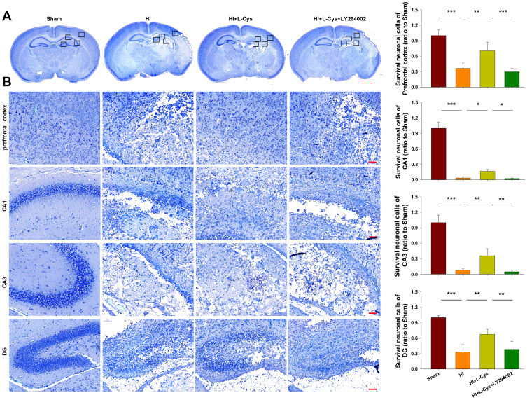
Figure 3.
Akt inhibition reverses the neuroprotective effects of L-Cysteine in HI. (A) Representative images of Nissl staining from each group at 72 h post-HI (Scale bar = 1000 µm). (B) Magnified views of boxed regions in A showing neuronal cell loss (Scale bar = 50 µm). Data are expressed as the number of surviving neuronal cells within each group relative to that of the Sham group within the different areas (N=5/group). Values represent the mean ± SD, *p < 0.05, **p < 0.01, ***p < 0.001 according to ANOVA.

