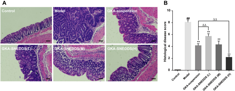
Figure 4.
(A) Representative H&E staining histological sections showing inflammatory changes in the colon. Scales bar, 100μm. (B) Histological disease score. The results were expressed as mean ± SD. ##p < 0.01 compared with the control group, **p < 0.01 compared with the model group, ΔΔp < 0.01 compared with the GKA-suspension-treated group.

