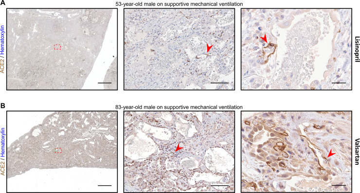
Fig 5. Two patients on ACEI/ARB therapy exhibit intense endothelial ACE2 expression.
A) Representative images of lung stained for ACE2 from a 53-year-old man with organizing diffuse alveolar damage superimposed on fibrosing interstitial lung disease are shown using a 1x objective (left, scale bar 3mm) with the region outlined by the red dashed box magnified at low power (middle, scale bar 200μm) and second field at high power (right, scale bar 50μm). AT2 cell hyperplasia can be seen with lower level ACE2 expression present along the alveolar septum (green arrowheads). Strongly stained endothelial cells (red arrowheads) can be seen throughout the entire specimen from this patient who was on daily lisinopril at the time of specimen collection. B) Representative images of lung stained for ACE2 from an 83-year-old man in the organizing phase of diffuse alveolar damage are shown using a 1x objective (left, scale bar 3mm) with the region outlined by the red dashed box magnified at low power (middle, scale bar 200μm) and second field at high power (right, scale bar 50μm). In addition to high ACE2 expression in reactive AT2 cells (green arrowheads), strong staining can be seen in numerous vascular endothelial cells (red arrowheads) throughout the thickened septal area in this patient on valsartan 4 days prior to specimen collection. Sections were stained for ACE2 using DAB and counterstained with hematoxylin.

