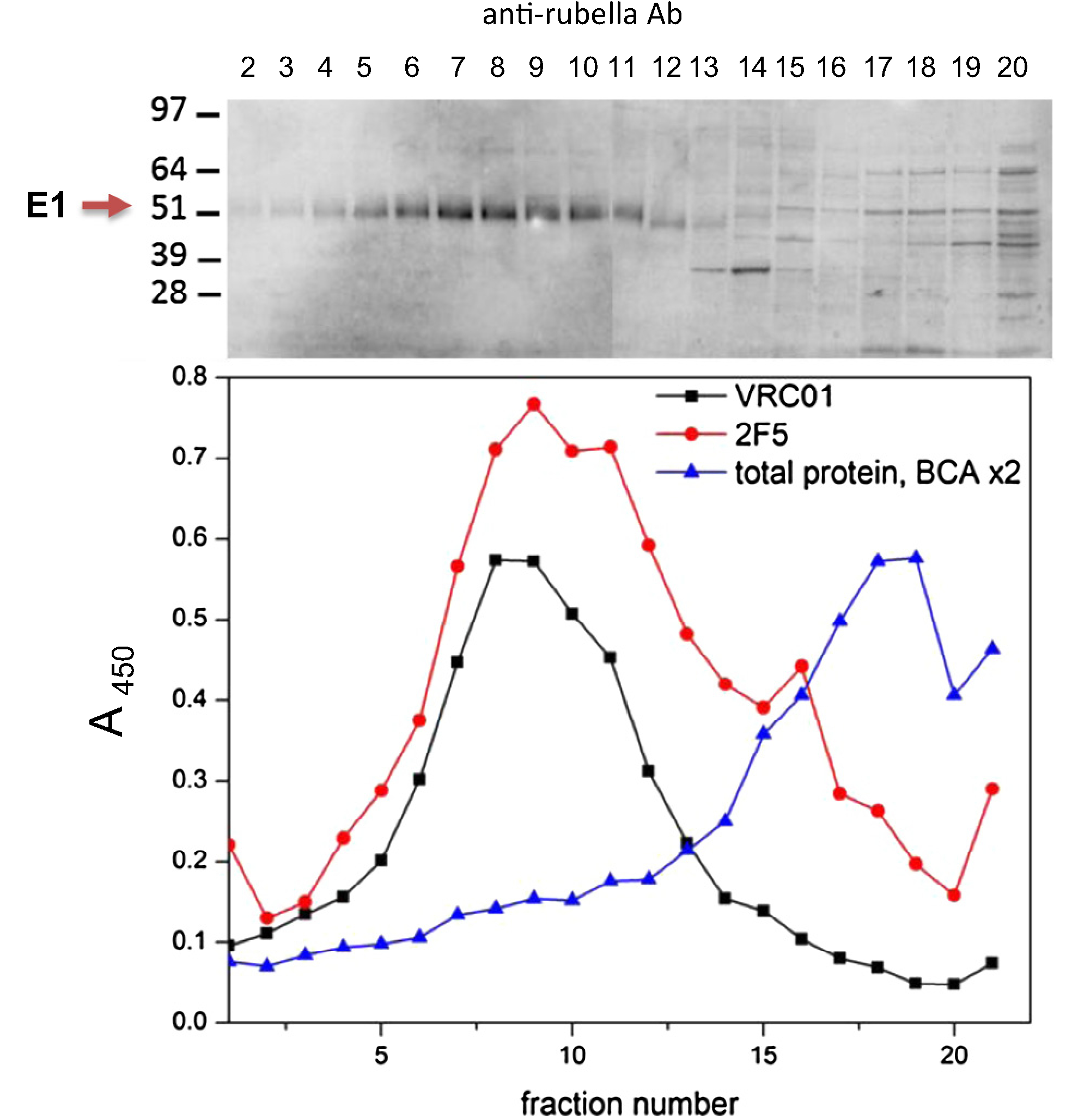
Fig. 3.

Sedimentation of rubella/eOD-GT6-min-gly virions on sucrose gradients. Cell lysates infected with eOD-GT6-min-gly vector were sedimented overnight on a 10–40% sucrose gradient. The rubella band was identified in peak fractions 5–11 by western blot with antibodies to rubella E1 protein (upper panel). The same fractions were assayed by ELISA (lower panel) with monoclonals VRC01 (black line) and 2F5 (red line). The eOD-GT6 peak coincides with the peak rubella fractions identified by western blot. Total protein content in the fractions is shown (blue line).
