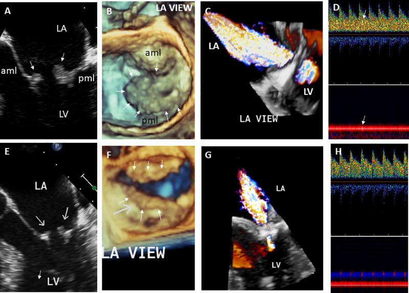
Fig 2. 29-year- old male with Libman-Sacks endocarditis complicated with acute stroke and cognitive dysfunction.

A. This two-dimensional (2-D) TEE view demonstrates large (area of 1.5 cm2), sessile, and oval shaped Libman-Sacks vegetations on the distal portions and atrial side of the anterior (aml) and posterior (pml) mitral leaflets (arrows). B. This 3-dimensional (3-D) TEE let atrial (LA) view of the mitral valve demonstrates large (area of 3.20 cm2) and protruding vegetations on the tip to mid portions and atrial side of the entire anterior and posterior mitral leaflets (arrows). C. Associated severe mitral regurgitation is demonstrated by 3D-TEE color Doppler. D. Transcranial Doppler of the middle cerebral artery demonstrates a microembolic event within the spectral Doppler (upper arrow) and within the vessel (lower arrow). On MRI, multiple small white matter lesions totaling a lesion load of 4.3 cm3 were demonstrated. His neurocognitive dysfunction was graded as severe. After 3 months of aspirin and clopidogrel, hydroxychloroquine, prednisone, and mycophenolate mofetil, mitral valve vegetations significantly decreased in size (arrows) by 2D (E) and 3D TEE (F) to vegetation areas of 0.22 cm2 and 0.79 cm2, respectively, and mitral valve regurgitation improved to mild (G). Also, cerebromicroembolism resolved (H), his brain lesion load decreased to 2.1 cm3, and his neurocognitive dysfunction improved to moderate degree. Abbreviations: LA = left atrium, LV = left ventricle, aml = anterior mitral leaflet, pml = posterior mitral leaflet.
