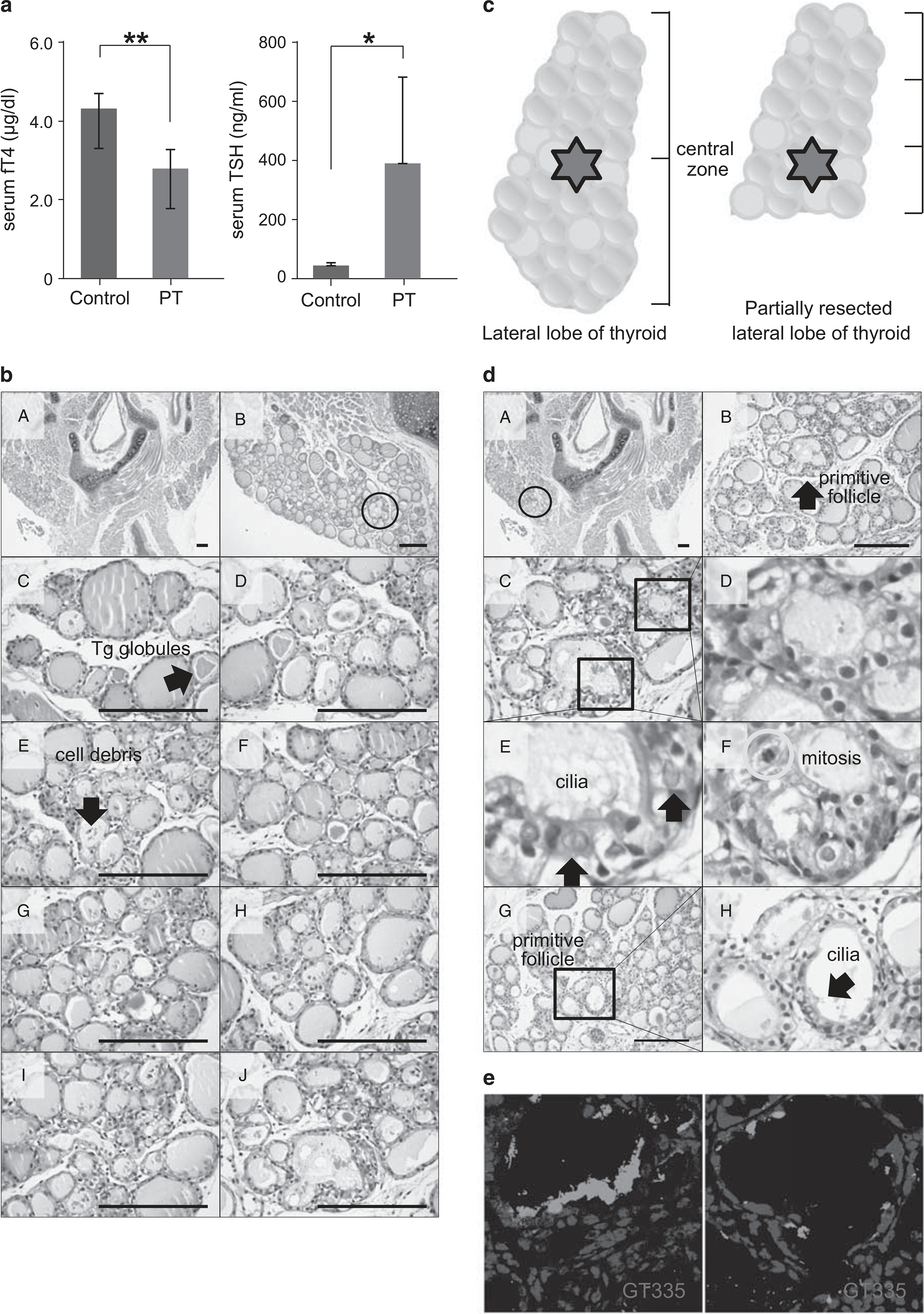
Figure 2.

Thyroid hormone change and histological analysis of thyroid sections at 10 days after partial thyroidectomy (PT). (a) The mean serum level of serum-free T4 (fT4) in PT was decreased to 2.74 ± 0.48 μg/dl at postoperative day 10 compared with 4.25 ± 0.37 μg/dl in the sham operation group (P = 0.0002). **P<0.01. The mean serum TSH level in PT was increased to 384.02 ± 286.96 ng/ml at postoperative day 10 compared with 43.49 ± 9.05 ng/ml in the sham operation group (P =0.031). *P<0.05. (b) Representative sequential images of the thyroid gland from the upper pole to the cutting margin. Hematoxylin and eosin (H&E) staining. Scale bar, 30 μm. (c) Schematic images show the central zone in the intact lateral lobe and the partially resected lateral lobe of the thyroid gland. (d) Representative images show regeneration centers of the thyroid gland. H&E staining. Scale bar, 30 μm. (e) Cilia in primitive follicular cells are immunoreactive to GT335, a monoclonal antibody directed against polyglutamylated tubulins.
