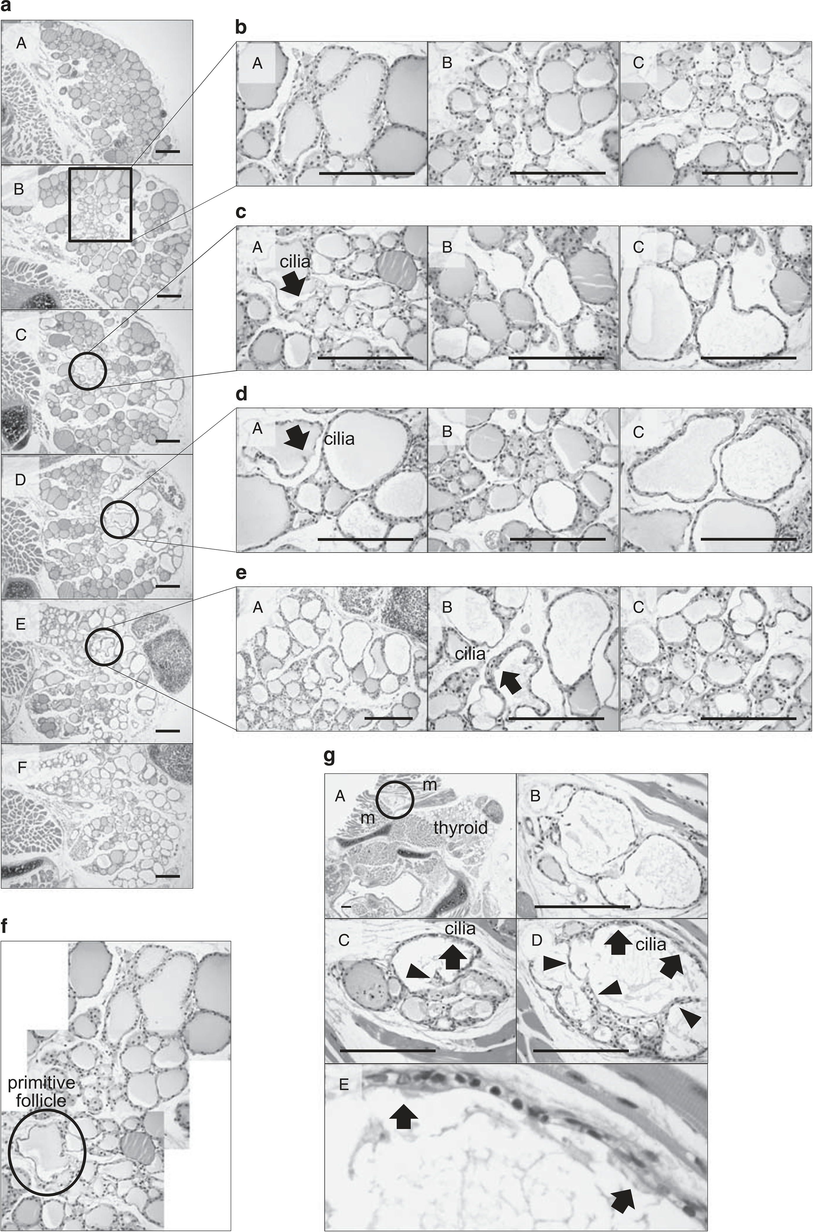
Figure 4.

Histological analysis of thyroid sections at 3 weeks after partial thyroidectomy. (a) Six representative serial sections are shown. The circle indicates the budding type of mother follicle-derived folliculogenesis. Boxed and circled regions are magnified in (b–e). (f) Reconstruction of follicles from serial sections is shown. (g) Lumen-dividing type of folliculogenesis is observed in the intramuscular spaces of the sternothyroid muscle. Arrow, ciliated primitive thyroid follicular epithelial cells; arrow head, papillary projections of a thyroid follicular epithelial cell. m, skeletal muscle. Hematoxylin and eosin (H&E) staining. Scale bar, 30 μm.
