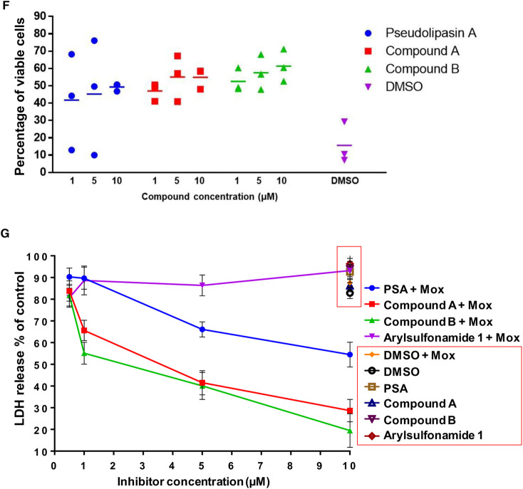
Figure 6. Compounds synergise with moxifloxacin to mitigate cell death induced by ExoU expressing PA103 over 24 h.
(A) Moxifloxacin (Mox) at the established MIC of 2 µM was added to scratched HCE-T cells that had been infected with PA103 ΔUT: ExoU at an MOI of 2.5 for 24 h. The number of CFU in the cell culture medium was then deduced. (B) LDH release from scratched HCE-T cells after 24 h infection in the presence of moxifloxacin at the MIC. (C) Live/Dead fluorescence microscopy analysis of scratched HCE-T cells 24 h post infection, without and with moxifloxacin at the MIC. (D) Live/Dead fluorescence microscopy analysis of scratched HCE-T cells 24 h post infection, in the presence of varying concentrations of indicated compound, with moxifloxacin present at the MIC. (E) Measurement of total scratch area (mm2) in compound treated HCE-T cells 24 h post infection in the presence of moxifloxacin. (F) Percentage of viable cells calculated within the scratch margin 24 h after infection in the presence of moxifloxacin. (G) LDH assay for dose response analysis of inhibitors analysing protective effect of compounds on scratched then infected HCE-T cells after 24 h incubation in the presence or absence of moxifloxacin at the MIC.


