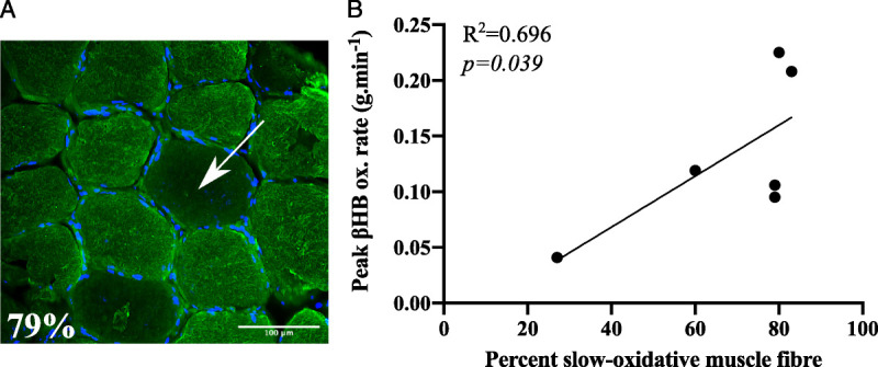
FIGURE 4.

Association between skeletal muscle fiber type and peak exogenous βHB oxidation rate. A, Vastus lateralis biopsy sample stained with an antislow skeletal myosin heavy-chain primary antibody (fluorescent green). Nuclei are represented in blue (stained with DAPI). Example nonstained fibers (representing fast-glycolytic (IIa) and fast (IIb) fibers) are indicated with the white arrow. B, Relationship between peak exogenous d-βHB oxidation rate (in grams per minute) and skeletal muscle phenotype (in percent).
