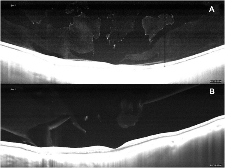
Figure 3.
(A) A PVD was identified by ultrasound, however the SS-OCT revealed adhesions of the vitreous at the fovea and the optic nerve head. The optically empty pockets seen within the vitreous of the SS-OCT image may represent a situation that is not easily identified with ultrasound. (B) SS-OCT revealed a small residual adhesion of the vitreous to the optic nerve head that was not identified by ultrasound.

