Figure 1.

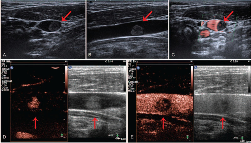
Ultrasonography: The image of the 2-dimensional ultrasound transverse section (A) and longitudinal section (B); (C) The image of the superb microvascular imaging; The images of the arterial phase (D) and the venous phase (E) of contrast-enhanced ultrasonography. The red arrows are pointing to the neoplasm.
