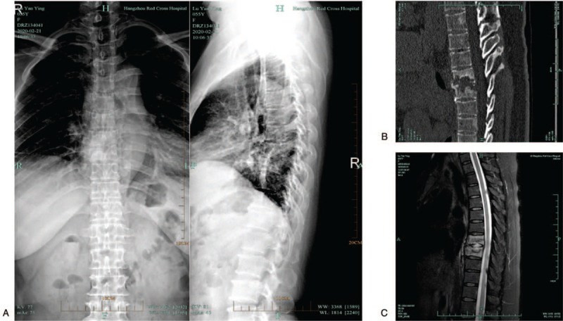
Figure 1.

Preoperative imaging data of patients with thoracic spine tuberculosis. (A) X-ray lateral view of the thoracolumbar spine, showing high density of T7 and T8 vertebral bodies, bone destruction, and narrowed intervertebral space. (B) It is a sagittal CT scan of the thoracic spine, showing the destruction of T7 and T8 vertebral bodies and the formation of cavities. (C) Sagittal radiographs of thoracic spine MRI fat-reducing images, showing high signal shadows of T7 and T8 vertebral bodies, and destruction of intervertebral discs. CT = computed tomography, MRI = magnetic resonance imaging.
