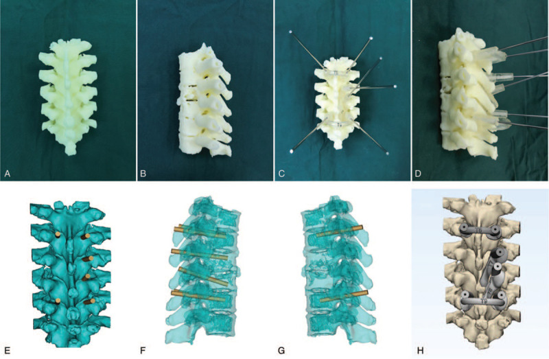
Figure 2.

Schematic diagram of the 3D printed model and surgical guide plate of patients with thoracic spine tuberculosis. (A, B) They are the front view and side view solid images of 3D printed diseased vertebral models accurately digitized according to medical imaging software. (C, D) Front and side views of the 3D navigation module implanted with nail channels. (E, F, G) It is the virtual front view, left side view, and right side view of the computer software simulating the 3D printing guide computer. (H) The computer accurately simulates the front view of the vertebral pedicle implanted after the diseased vertebral body. 3D = 3-dimensional.
