
Figure 4.
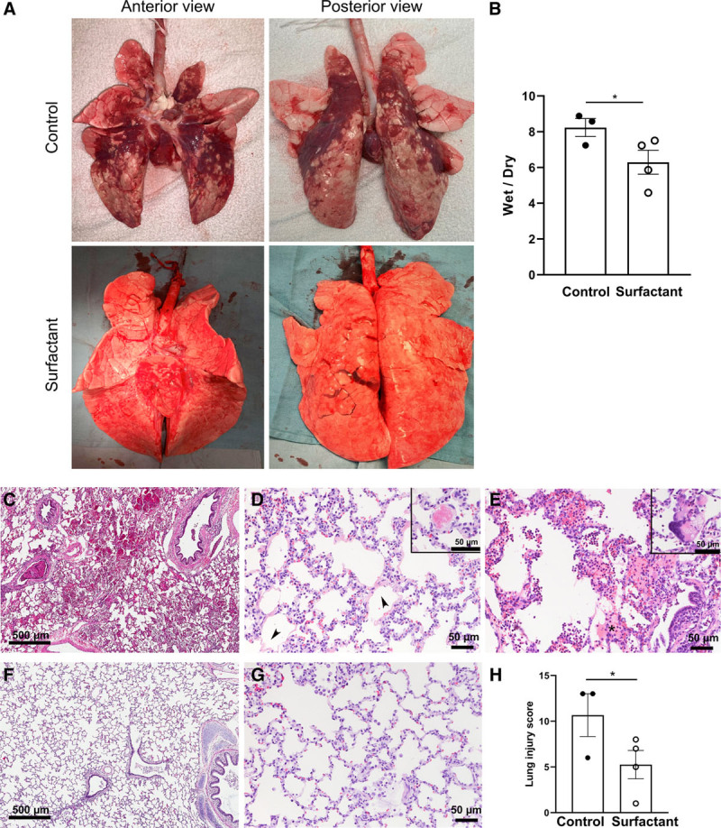
Macro- and microscopic appearance of lung tissue and cytokine levels. A, Gross appearance of the lungs is shown while being ventilated ex vivo with peak inspiratory pressures of 20 cm H2O. Compared with the well-inflated lungs of the surfactant-treated animals, the control lungs are collapsed with patchy hemorrhage. B, The wet-to-dry weight ratio was lower in Surfactant group than Control group. Representative lung histology in control (C–E) and surfactant-treated animals (F, G). C, Hemorrhage, (D) septal thickening, hyaline membranes (arrowheads), (E) neutrophilic inflammation and proteinaceous exudate (asterisk) were more prominent in the control lungs; vascular thrombi (D, inset) and multinucleated giant cells (E, inset) were occasionally seen. The alveolar architecture was generally well preserved in the surfactant-treated group with less inflammation and thin alveolar walls (F and G). H, The Lung Injury Score was lower in surfactant-treated than control animals. Values are means ± sem. *p < 0.05.