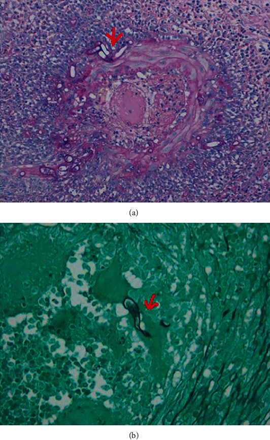
Figure 12.

A small intestine biopsy specimen shows the Splendore-Hoeppli phenomenon in a patient diagnosed with colonic Basidiobolomycosis ((a) red arrow). Grocott stain of the small intestine shows Basidiobolus hyphae (red arrow), which appears thin-walled, septated hyphae surrounded by eosinophilic material (b). (Images obtained with permission from Kurteva et al. [81]).
