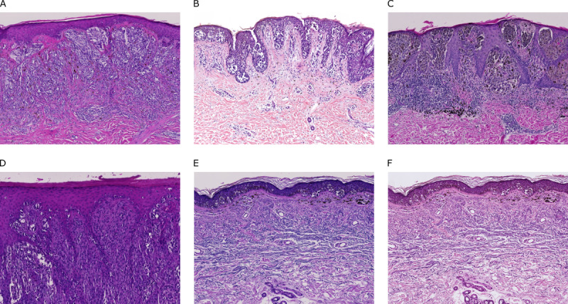
Figure 1.

Comparison of exemplary whole slide image sections obtained at different institutes: (A) Institute of Pathology, University Hospital Heidelberg, University of Heidelberg, Heidelberg, Germany (Zeiss scanner; Carl Zeiss AG); (B) Department of Dermatology, University Hospital Kiel, University of Kiel, Kiel, Germany (3DHISTECH scanner; 3DHISTECH Ltd); (C) Private Institute of Dermatopathology, Mönchhofstraße 52, Heidelberg, Germany (Zeiss scanner); (D) Department of Dermatology, University Medical Center Mannheim, University of Heidelberg, Mannheim, Germany (Zeiss scanner); and (E) Private Institute of Dermatopathology, Siemensstraße 6/1, Friedrichshafen, Germany (Zeiss scanner). (F) The same slide section as shown in (E) but scanned with a Hamamatsu scanner (Hamamatsu Photonics KK) rather than a Zeiss scanner.
