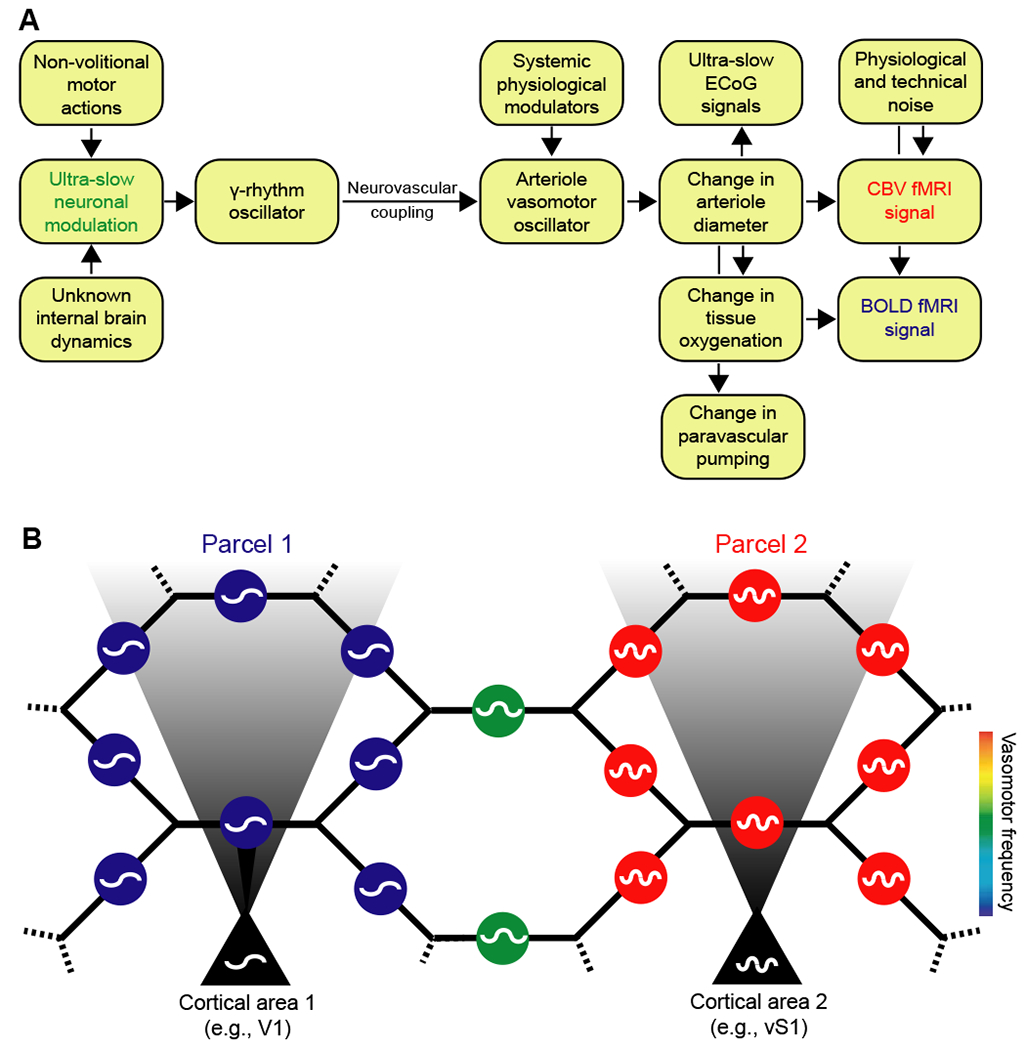
Figure 8. Coupled oscillator model for resting-state neurovascular coupling.

(A) Interactions for one region. Variations in γ-band electrical power lead to partial entrainment of the vasomotor oscillations in the smooth muscle of cortical surface and penetrating arterioles. Increases in γ-band electrical power dilate the arterioles and lead to an increase in the supply of fresh blood, as measured by CBV fMRI. The increase in diameter subsequently leads to an increase in blood oxygenation and a positive change in the BOLD fMRI signal. Coupling can be via callosal projections or via common input.
(B) Circuit of the pial arteriole network in terms of coupled smooth muscle oscillators. Each oscillator represents the activity of all muscles in one arteriole. The oscillators communicate with one another via the hexagonal lattice of electrically-joined endothelial cells that form the lumen (Fig. 6). Oscillators in a given region of the brain may also receive electrical input from underlying neuronal oscillators. This input can dominate and lead to a parcellation of the frequency of vasomotor oscillations, as observed by fMRI (Fig. 2B). Vasomotion is synchronous within a given region of the brain yet can occur with different frequencies in different regions; e.g., labeled blue and red for regions 1 and 2, respectively, and labeled green for an intermediate region without neuronal drive.
