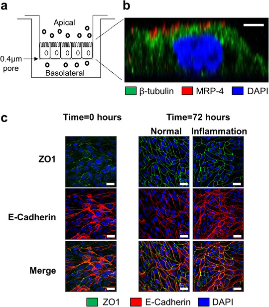
FIGURE 1.

Establishment of a Transwell model to examine polarized sEV production by human primary PTEC. (a) Human primary PTEC were seeded onto Transwell inserts (0.4 μm pore size) and grown to confluence. The defined medium (DM) was subsequently exchanged with fresh DM for normal control PTEC and fresh DM supplemented with 100 ng/ml IFN‐γ and 20 ng/ml TNF‐α for inflammatory PTEC and then further cultured for 72 h. PTEC culture medium was then harvested from the upper (apical) compartment and lower (basolateral) compartment for downstream sEV isolation. (b) Immunofluorescent microscopy of inflammatory PTEC monolayers on Transwell inserts (lateral projection) stained for β‐tubulin (green), MRP‐4 (red) and DAPI (blue). Scale bar represents 2 μm. One representative of three PTEC donor experiments. Equivalent staining profile was observed for normal control PTEC monolayers. (c) Immunofluorescent staining of PTEC monolayers on Transwell inserts for ZO1 (green), E‐Cadherin (red) and DAPI (blue) at time = 0 h and following 72 h culture under normal and inflammatory conditions. Scale bars represents 20 μm. One representative of two PTEC donor experiments.
