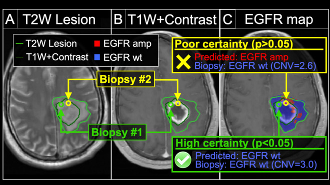
Figure 4.
Model uncertainty can inform the likelihood of achieving an accurate radiogenomics prediction for EGFR amplification status. We obtained two separate biopsies (#1 and #2) from the same tumor in a patient with primary GBM. The (A) T2W and (B) T1+C images demonstrate the enhancing (dark green outline, T1W+Contrast) and peripheral non-enhancing tumor segments (light green outline, T2W lesion). The (C) radiogenomics color map shows regions of predicted EGFR amplification (amp, red) and EGFR wildtype (wt, blue) status overlaid on the T1+C images. For biopsy #1 (green circle), the radiogenomics model predicted EGFR wildtype status (blue) with high certainty, which was concordant with biopsy results (green box). For biopsy #2 (yellow circle), the radiogenomics model showed poor certainty (i.e., high uncertainty), with resulting discordance between predicted (red) and actual EGFR status (yellow box).

