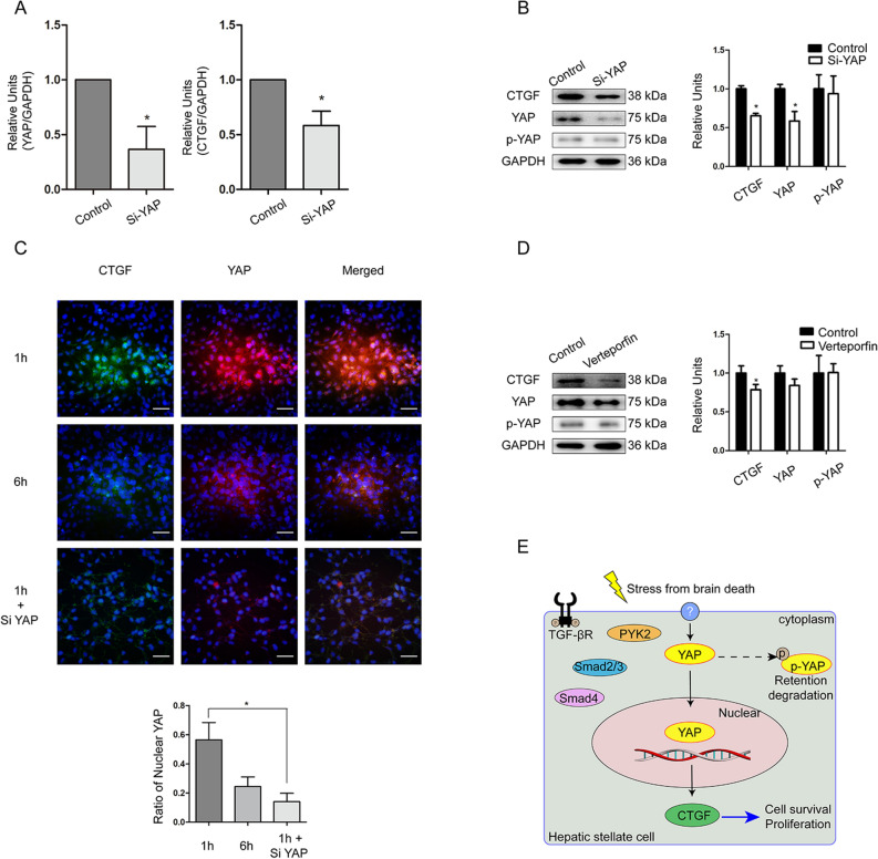
Figure 4.
Regulation of CTGF through YAP in LX2 cells treated with Si-RNA or YAP inhibitor Verteporfin under hypoxia for 3 h and reoxygenation for 1 h. LX2 cells treated with vehicle of Si-RNA or Verteporfin were regarded as control. (A) Changes of YAP and CTGF mRNAs in LX2 cells after YAP-siRNA treatment under hypoxia/reoxygenation determined by RT-qPCR (*P < 0.05 between siRNA and control). (B) Changes of CTGF, YAP, and p-YAP protein levels after YAP-siRNA treatment in LX2 cells determined by WB (*P < 0.05 between siRNA and control). (C) Immunofluorescence staining of CTGF (green) and YAP (red) in LX2 cells under hypoxia/reoxygenation; DAPI (blue) was used for nuclear staining (scale bar = 20 μm, ×200 magnification) and semi-quantitation of nuclear YAP (*P < 0.05). (D) Changes of CTGF, YAP, and p-YAP expression in LX2 cells after treatment of the YAP inhibitor Verteporfin under hypoxia/reoxygenation (*P < 0.05). (E) Schematic of the proposed mechanism of CTGF regulation in the non-steatotic liver after brain death.

