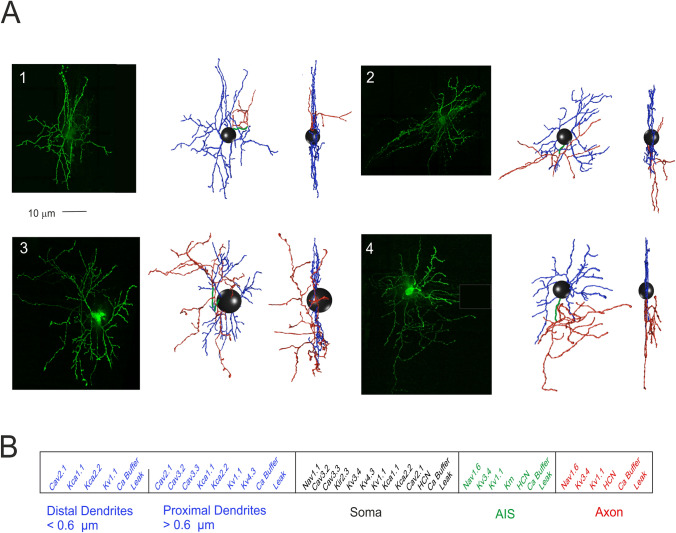
Figure 1.
Stellate cell morphological reconstruction. (A) Confocal microscopy imaging of four mouse SCs filled with Lucifer yellow (scale bar 10 μm) are shown along with the corresponding digital reconstruction with Neurolucida (visualization with Vaa3D simulator; right). The 3D morphological reconstructions of SCs include dendrites (blue), soma (black), AIS (green) and axon (red). (B) The SC model is divided into five electrotonic compartments and endowed with specific ionic mechanisms according to immunohistochemical data. The proximal and distal dendrites are distinguished at the cut-off diameter of 0.6 µm. Ionic channels include Na+, K+ and Ca2+ channels and a Ca2+ buffering system.

