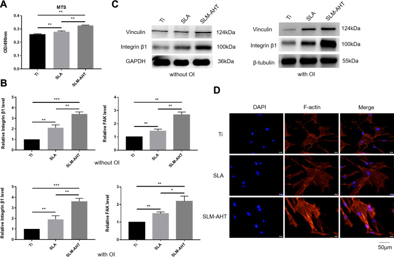
Figure 2.
PDLSCs adhesion and actin cytoskeletal state on different titanium surfaces. (A) MTS assays for cell early adhesion on different titanium surfaces after 2 hours of culture. (B) The relative mRNA levels of integrin β1 and FAK in PDLSCs cultured on Ti, SLA and SLM-AHT surfaces for 3 days with and without osteoinduction. (C) The total protein expression of integrin β1 and vinculin in PDLSCs cultured on different surfaces for 3 days with and without osteoinduction. (D) Immunofluorescence staining assays for actin cytoskeleton state of PDLSCs cultured on different surfaces for 3 days (red, F-actin; blue, DAPI). Error bars represent SD (n = 3). *P <0.05,**P < 0.01,***P < 0.001.

