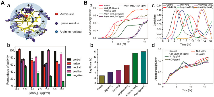
Figure 6.
(A(a)) Structure of β-lactamase. The active site (red) of β-lactamase is surrounded by lysine and arginine (blue) residues. (b) Activity of β-lactamase in the presence of increasing concentration of various 2D MoS2 in HEPES buffer (pH 7.3) using nitrocefin as a substrate. (B) Growth curve analysis for the bacterial strain MRSA, treated with ampicillin with various concentrations of negative functionalized MoS2 at 37°C. (a) The bacterial growth curves over a period of 16 h, in a real-time kinetic cycle. (b) The lag phase diagram and (c) first derivative growth rate for quantitative analysis of the growth curve. (d) Growth curve analysis for the bacterial strain MRSA, treated with various concentrations of negative ligand molecule at 37°C over a period of 16 h, in a real-time kinetic cycle. Reprinted with permission from Ali SR, Pandit S, De M. 2D-MoS2-Based β-Lactamase Inhibitor for combination therapy against drug-resistant bacteria. ACS Applied Bio Materials. 2018;1(4):967–974. Copyright (2018) American Chemical Society.37

