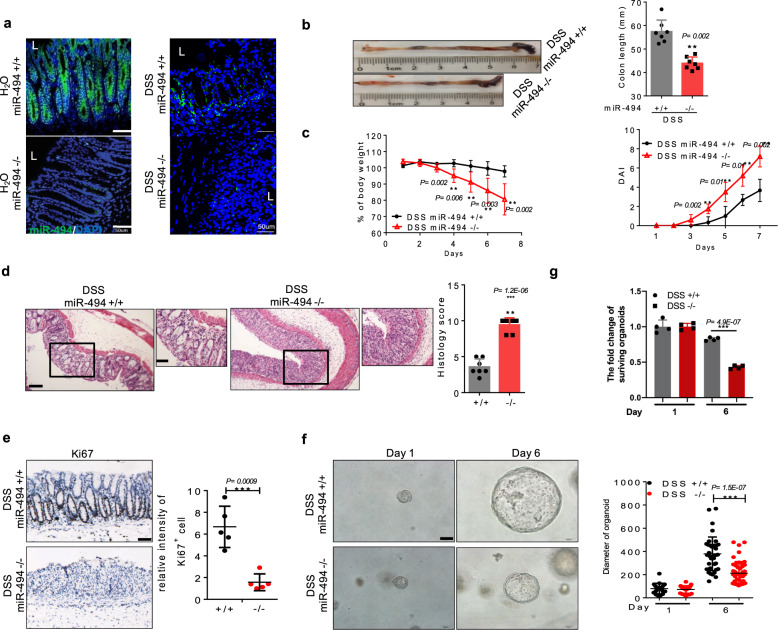
Fig. 2. miR-494−/− mice exhibit abnormalities in crypt stem cell identity in DSS-induced colitis.
a–g miR-494 knockout mice (miR-494−/−, −/−) and wide-type mice (WT, miR-494+/+, +/+) at 8-weeks old were provided with free access to drinking water with 3% DSS for 7 days. Day 0 is the initiation of DSS treatment. a Representative images of miR-494-3p expression in colon tissue from RNA-FISH assay, scale bar: 50 μm. b Typical images of the colon from miR-494−/− mice and WT mice on day 7. Colon length was measured on day 7 after killing. c Body weight changes and DAI. d Hematoxylin and eosin (H&E) images of colon tissue and histology score (0–12), scale bar: 200 μm. The scale bar of close-up image is 100 μm. e IHC was performed for the proliferation marker Ki67 in colon, scale bar: 100 μm. f Representative images of colon organoids derived from colonic crypt that were isolated from DSS-treated mice (on day 7) and then cultured in vitro for 6 days (right), scale bar: 100 μm. e, f The diameter of organoids and number of Ki67+ cells were measured by ImageJ. g The quantification of surviving organoids. Scale bar: 100 μm. Data are presented as mean ± S.D. and analyzed by non-paired two-tailed Student’s t-test in panels (b–e). ***P ≤ 0.001, **P ≤ 0.01, *P ≤ 0.05.

