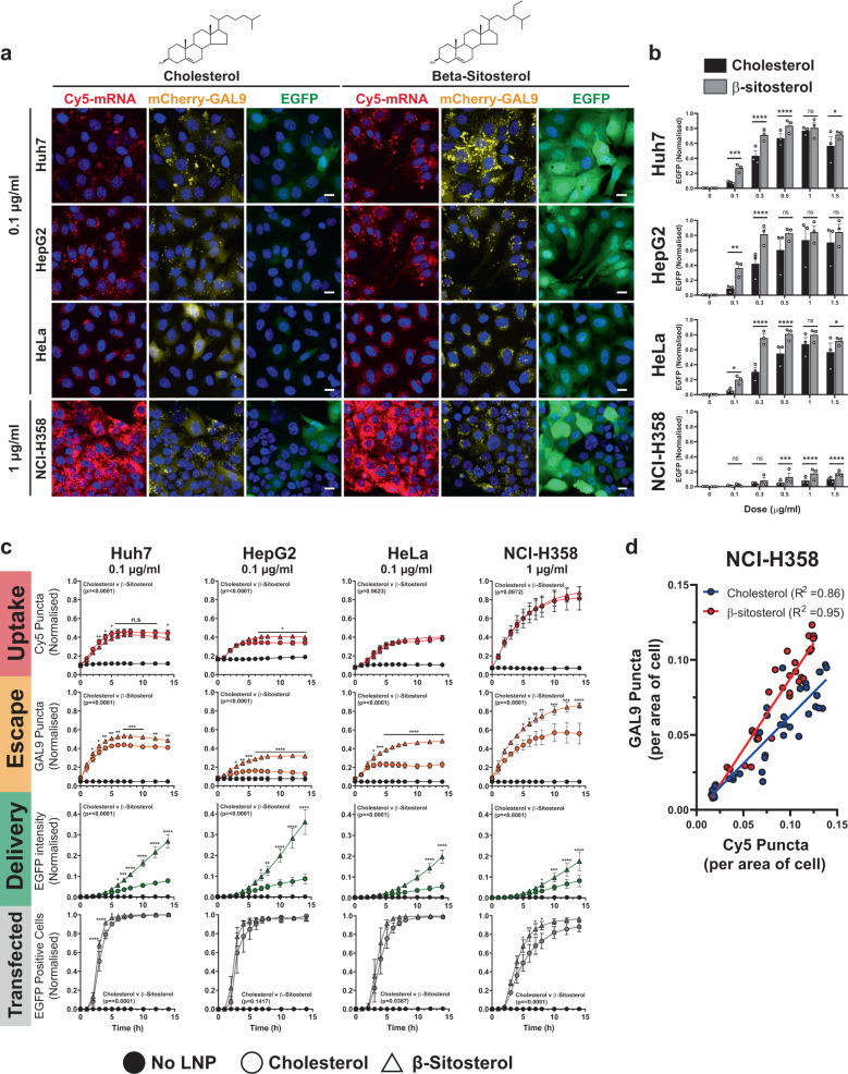
Fig. 5. β-Sitosterol modifies endosomal escape rate.
a Huh7, HepG2, HeLa and NCI-H358 mCherry-GAL9 cells were dosed at indicated concentrations of MC3-LNPs formulated with cholesterol or β-sitosterol. Images are representative of 14 h post-dosing. Scale bar = 20 µm. b Comparison of cell EGFP fluorescent intensity at 14 h after incubation with Cholesterol or β-sitosterol particles in Huh7, HeLa, HepG2 and NCI-H358 cells across 0.1–1.5 µg/ml doses. Values represent normalised EGFP intensity ± SEM from n = 3 independent experiments. c Quantitation of (a) examining the formation of Cy5 positive structures, mCherry-GAL9 structures and EGFP fluorescence per cell over time. Values represent normalised means ± SEM from n = 3 independent experiments. Significance was determined in (b, c) for full 0–14 h time courses by two-way ANOVA followed by Tukey’s multiple comparison test where *p < 0.05, **p < 0.01 ***p < 0.001, ****p < 0.0001 and ns not significant. d Comparison of total Cy5 or GAL9 puncta over time obtained from (c) for cholesterol or β-sitosterol particles in NCI-H358 cells. Linear regression carried out and R2 values displayed on the graph.

