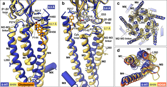
Fig. 6. Comparison of serotonin-bound 5HT3R-Salipro and 6HIN structures.
a, b Superposition of the serotonin-bound 5HT3R-Salipro (chain C) and 5HT3R-detergent (6HIN)17 structures at a the cholesterol-binding pocket viewed from the membrane (M3 is omitted for clarity) or b at the Cys loop, β1-β2 loop, M2–M3 linker junction viewed from the neighbouring subunit (M1 and part of the Cys loop and TMD are omitted for clarity). Distances are labelled in the same colour scheme as the structures. Density sharpened using RELION with auto-determined b-factor, shown in transparent grey for residues R218 and E53, at σ 6.5. c, d Top views of the TMD, with d including the 5HT3R-detergent 6DG8 structure and showing single subunits only for clarity.

