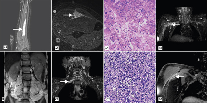
Figure 2.
Peripheral nerve tumors - (A1) Postcontrast T1 weighted fat saturated coronal image of left forearm, (A2) STIR (short T1 inversion recovery) axial image at the level of left mid forearm showing fusiform enlargement with preserved fascicular pattern, intense STIR hyperintensity and post contrast enhancement of the left ulnar nerve (arrows) in a patient of perineuroma. (A3) Semithin section, toluidine blue stain of fascicular biopsy of left ulnar nerve showing pseudo-onion bulb appearance, which stained positive for epithelial membrane antigen and S-100 (not shown) suggestive of perineuroma (40×). (B) T2 weighted coronal image of the lumbosacral plexus showing plexiform neurofibromas along the lumbosacral plexuses. (C1) Reconstructed STIR MIP (maximum intensity projection) coronal image of the brachial plexus shows a small schwannoma along the post ganglionic right C7 root (arrow). (C2) Hematoxylin and eosin stain showing palisading appearance suggestive of schwannoma (40×). (D1) Reconstructed STIR MIP coronal and (D2) postcontrast T1 weighted fat saturated images of the right brachial plexus showing an ill-defined intensely enhancing lesion along the cords of right brachial plexus (arrows) which turned out to be hemangioendothelioma on histopathological examination

