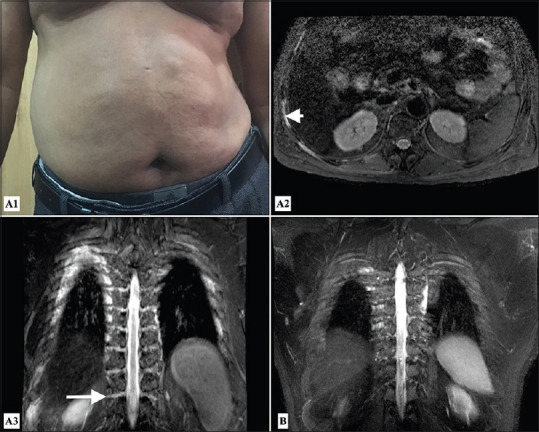
Figure 3.

Diabetic truncal neuropathy - Clinical image (A1) showing protruded abdomen on right side due to weakness of abdominal wall muscles; MR images (A2) STIR axial at the level of D12 vertebra and (A3) STIR MIP coronal image of the dorsal spine showing thickening and hyperintense signal of the right sided dorsal nerve roots (arrow) and right intercostals nerves (arrowhead) in a patient with diabetic dorsal truncal neuropathy as opposed to (B) normal STIR MIP coronal image of the intercostal nerves for comparison
