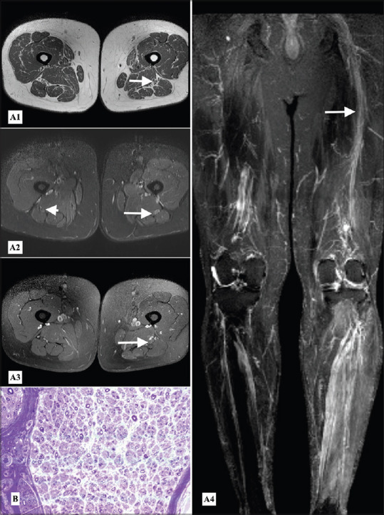
Figure 4.

Sciatic mononeuropathy - MR Neurography (A1) T1weighted axial, (A2) STIR axial, (A3) postcontrast T1 weighted axial images at the level of mid-thigh and (A4) STIR MIP reconstructed coronal image of the lower limbs showing mild diffuse thickening, abnormal STIR hyperintense signal and postcontrast enhancement of the left sciatic nerve (arrows) as compared to normal appearing right sciatic nerve (arrowhead), (B) Semithin section, toluidine blue stain of fascicular biopsy of left sciatic nerve showing axon loss and regenerating fibers, preserved myelin and no evidence of vasculitis, granuloma or malignancy (40×)
